

Médiathèque SET

L'intégration de la NMES dans le parcours postopératoire du LCA

TENS: Much More Than a “Pain Signal Blocker” TENS is a familiar tool for most healthcare professionals. Accessible, non-pharmacological, and relatively easy to use, it has long been part of the therapeutic options available for pain management. Yet its effectiveness remains a topic of debate. While some patients report meaningful relief, others experience little or no benefit. Does TENS truly work for complex chronic pain conditions? The answer is more nuanced than a simple yes or no. To understand why, we need to move beyond the traditional view of TENS as a device that merely "blocks" or temporarily disrupts pain signals. This concept originates from the well-known Gate Control Theory of pain. By stimulating the sensory nerve fibres responsible for touch and vibration, TENS activates inhibitory mechanisms within the spinal cord. In essence, it helps "close the gate" on certain pain messages before they reach the brain (Martimbianco et al., 2019; Smith et al., 2023). However, decades of research have demonstrated that its effects extend well beyond this mechanism. Indeed, TENS may influence pain modulation at multiple levels of the nervous system. Near the painful region, it can affect mechanisms involved in pain sensitivity. Within the spinal cord, it may reduce the transmission of nociceptive signals. It can also activate endogenous pain-control pathways within the brain itself (Gibson et al., 2019; Smith et al., 2023). In other words, the effects of TENS are not confined to the area beneath the electrodes. This broader perspective becomes particularly relevant in conditions such as fibromyalgia, complex regional pain syndrome (CRPS), and certain forms of neuropathic pain. In these disorders, pain is not driven exclusively by tissue pathology; it is also influenced by the way the nervous system processes and regulates sensory information. This distinction fundamentally changes how we think about the clinical role of TENS. Central Sensitization: When the Nervous System Turns Up the Volume To appreciate the potential role of TENS in complex pain conditions, it is helpful to understand the concept of central sensitization. Although the term may sound technical, the underlying idea is straightforward: the nervous system becomes increasingly responsive to incoming signals and amplifies the perception of pain. A useful analogy is that of a stereo system with the volume turned up much higher than intended. As a result, stimuli that would normally be painless may become painful, a phenomenon known as allodynia. Likewise, stimuli that are already painful may produce a disproportionately intense response, referred to as hyperalgesia. These changes may also be accompanied by impairments in the body's natural pain-inhibitory systems. In some individuals living with persistent pain, the brain's internal "braking mechanisms" appear less effective at dampening painful inputs (Dailey et al., 2013). Central sensitization plays a particularly important role in fibromyalgia. It may also contribute, to varying degrees and alongside other mechanisms, to certain presentations of CRPS and neuropathic pain. Nevertheless, it would be overly simplistic to attribute all of these conditions solely to central sensitization. This is where the rationale for using TENS becomes especially compelling. In individuals with fibromyalgia, Dailey et al. (2013) observed reductions in pain and hypersensitivity during TENS application. Notably, some changes were detected at sites distant from the area being stimulated, suggesting that TENS may alter how the nervous system regulates pain rather than simply producing a localized effect beneath the electrodes. Fibromyalgia: The Evidence Is Becoming Increasingly Difficult to Ignore Among the complex pain conditions discussed in this article, fibromyalgia likely has the strongest body of evidence supporting the use of TENS. Early studies showed that even a single TENS session could reduce pain, fatigue, and hypersensitivity in individuals with fibromyalgia (Dailey et al., 2013). Researchers subsequently shifted their attention toward a more practical question: Can reducing pain help people move more effectively? A randomized controlled trial published in 2020 found that home-based TENS used during activity significantly reduced movement-evoked pain and fatigue compared with placebo stimulation (Dailey et al., 2020). A systematic review and meta-analysis published in 2023 further highlighted the importance of considering TENS dosage when evaluating treatment outcomes in fibromyalgia (Amer-Cuenca et al., 2023). More recently, Dailey et al. (2026) assessed the addition of TENS to physiotherapy across 28 outpatient clinics. Their findings demonstrated clinically meaningful reductions in movement-related pain, along with improvements in fatigue and overall disease impact. These benefits appeared rapidly and were maintained through the six-month follow-up period. Why are these findings particularly important? Because the goal of treatment is not necessarily complete pain elimination. If TENS reduces pain enough to allow patients to walk farther, perform their exercises, or participate more actively in rehabilitation, its clinical value becomes far more significant. In this context, TENS evolves from a modality aimed solely at symptom relief into a tool that actively facilitates function and participation. CRPS: A Plausible Mechanism Does Not Guarantee Clinical Effectiveness Complex Regional Pain Syndrome provides an excellent example of a key principle in evidence-based practice: a treatment that makes physiological sense is not automatically effective in the clinical setting. CRPS is a multifactorial condition in which numerous mechanisms may contribute to pain and disability. Alterations involving the nervous system, inflammation, cutaneous sensitivity, motor control, and central pain processing may all play a role (Ferraro et al., 2023, 2024). Given these mechanisms, there is a reasonable rationale for considering TENS in some patients. However, the clinical evidence remains limited. Available studies are relatively few in number and generally include small sample sizes. Consequently, systematic reviews continue to report substantial uncertainty regarding the independent effectiveness of TENS for reducing pain and disability in CRPS (Smart et al., 2022). For this reason, TENS should not be promoted as a treatment for CRPS itself. A more appropriate role may be as an adjunct to rehabilitation, particularly when it helps patients tolerate activities that would otherwise be difficult to perform. Depending on the clinical presentation, management may include pain education, graded exposure to movement, sensory retraining, and various sensorimotor interventions (Ferraro et al., 2024; Lloyd et al., 2021). In practice, TENS may support CRPS rehabilitation, but it is not a substitute for a comprehensive treatment approach. Neuropathic Pain: The Right Question Isn't Simply “Does It Work?” Although the question appears straightforward, the answer is anything but. The term neuropathic pain encompasses a broad range of conditions with distinct underlying mechanisms. Diabetic peripheral neuropathy, for example, differs substantially from neuropathic pain resulting from a spinal cord injury, and these conditions may not respond to treatment in the same way. This distinction is highlighted in a recent meta-analysis. Overall, the effect of TENS on neuropathic pain was modest and failed to reach statistical significance. However, when investigators examined specific pain conditions, important differences emerged. No clear benefit was observed in diabetic neuropathy, whereas more favourable outcomes were reported in individuals with neuropathic pain associated with spinal cord injury (ElMeligie et al., 2025). These findings should be interpreted cautiously given the variability and overall quality of the available studies. Nevertheless, they emphasize an important clinical principle: Neuropathic pain is not a single entity, and it should not be approached as one when considering a trial of TENS. Rather than asking whether TENS works for neuropathic pain in general, a more meaningful question is: Which patient, with which type of pain, and for what clinical goal, is most likely to benefit from TENS? Why Doesn't TENS Produce the Same Results for Everyone? One of the main reasons the literature on TENS appears inconsistent is that not all TENS interventions are equivalent. Treatment intensity may vary. Session duration may differ. Electrode placement is often inconsistent across studies. Some patients use TENS while resting, whereas others use it during physical activity. Frequency settings and daily treatment exposure also vary considerably. As a result, two studies may both evaluate "TENS" while examining significantly different interventions. 1. Intensity Matters Evidence consistently suggests that conventional TENS should be delivered at an intensity that is strong, yet comfortable and non-painful. In clinical practice, there is often a tendency to reduce intensity excessively in an effort to avoid discomfort. However, stimulation that is barely perceptible may not provide an adequate therapeutic dose. While stronger is not always better, stimulation that is too weak may limit effectiveness. 2. High Frequency Versus Low Frequency: Only One Piece of the Puzzle Most TENS devices allow clinicians to adjust stimulation frequency. Higher frequencies are commonly used to produce a comfortable sensory sensation, whereas lower-frequency protocols may engage different pain-modulating mechanisms and often require different intensity levels.However, focusing exclusively on frequency may be misleading. There is unlikely to be a single "optimal" frequency that works for everyone. Instead, frequency should be considered alongside treatment intensity, duration of use, electrode placement, and, most importantly, the intended clinical outcome. 3. Timing May Be Just as Important as the Settings An often-overlooked finding from successful fibromyalgia trials is that TENS was frequently applied during activities known to provoke symptoms (Dailey et al., 2020; Dailey et al., 2026). This detail may be more important than it first appears. If the goal is to reduce movement-evoked pain, assessing TENS only while the patient is at rest may underestimate its clinical value. For many patients, TENS may be most useful when applied during walking, exercise, or other functional activities. In this context, the objective shifts from eliminating pain to making movement more tolerable and participation more achievable. 4. Electrode Placement Still Matters Electrodes are typically positioned over or near the painful area, or according to a clinically relevant anatomical distribution. In individuals with complex pain conditions and marked hypersensitivity, comfort and tolerability become critical considerations. Consequently, there is rarely a single electrode placement strategy that is appropriate for every patient presenting with the same diagnosis. Clinical reasoning and patient response should guide positioning decisions. 5. Why Not Test the Response First? This concept is particularly relevant in fibromyalgia. Vance and colleagues (2021) demonstrated that changes in movement-evoked pain and fatigue during an initial 30-minute TENS session were predictive of longer-term treatment response. From a clinical perspective, this supports a simple approach: Try it. Measure the response. Reassess. Rather than basing the decision solely on diagnosis, clinicians can use an individualized trial to determine whether TENS provides meaningful benefits for a specific patient. Could the Real Value of TENS Lie Elsewhere? TENS is neither a universal solution nor merely a device that blocks pain signals. Its value lies in how it is integrated into a broader management strategy: with the right patient, appropriate treatment parameters, and a clearly defined clinical objective. The evidence is particularly encouraging in fibromyalgia, whereas findings remain less convincing in CRPS and vary considerably across different neuropathic pain conditions. One conclusion, however, remains consistent: diagnosis alone is a poor predictor of treatment success. Dosage, context of use, and individual responsiveness all play a critical role. Importantly, the goal of treatment is not always complete pain relief. If TENS reduces pain sufficiently to facilitate movement, support exercise participation, or improve engagement in meaningful activities, it can become a valuable adjunct to rehabilitation. Ultimately, perhaps the most useful question is no longer: "Does TENS work?"but rather: "How can TENS be used to provide meaningful benefit for this particular patient?" References Amer-Cuenca, J. J., Badenes-Ribera, L., Biviá-Roig, G., Arguisuelas, M. D., Suso-Martí, L., & Lisón, J. F. (2023). The dose-dependent effects of transcutaneous electrical nerve stimulation for pain relief in individuals with fibromyalgia: A systematic review and meta-analysis. Pain, 164(8), 1645–1657. https://doi.org/10.1097/j.pain.0000000000002876 Dailey, D. L., Rakel, B. A., Vance, C. G. T., Liebano, R. E., Amrit, A. S., Bush, H. M., Lee, K. S., Lee, J. E., & Sluka, K. A. (2013). Transcutaneous electrical nerve stimulation reduces pain, fatigue and hyperalgesia while restoring central inhibition in primary fibromyalgia. Pain, 154(11), 2554–2562. https://doi.org/10.1016/j.pain.2013.07.043 Dailey, D. L., Vance, C. G. T., Rakel, B. A., Zimmerman, M. B., Embree, J., Merriwether, E. N., Geasland, K. M., Chimenti, R., Williams, J. M., Golchha, M., Crofford, L. J., & Sluka, K. A. (2020). Transcutaneous electrical nerve stimulation reduces movement-evoked pain and fatigue: A randomized, controlled trial. Arthritis & Rheumatology, 72(5), 824–836. https://doi.org/10.1002/art.41170 Dailey, D. L., Vance, C. G. T., Van Gorp, B. J., et al. (2026). Transcutaneous electrical nerve stimulation and pain with movement in people with fibromyalgia: A cluster randomized clinical trial. JAMA Network Open, 9(3), e262450. https://doi.org/10.1001/jamanetworkopen.2026.2450 ElMeligie, M. M., Gomaa, Y. S., Taha, E., et al. (2025). Neuropathic pain relief through transcutaneous electrical neuromuscular stimulation: Insights from a systematic review and meta-analysis of clinical evidence. BioMed Research International, 2025, 5328365. https://doi.org/10.1155/bmri/5328365 Ferraro, M. C., Cashin, A. G., Wand, B. M., et al. (2023). Interventions for treating pain and disability in adults with complex regional pain syndrome: An overview of systematic reviews. Cochrane Database of Systematic Reviews, 2023(6), CD009416. https://doi.org/10.1002/14651858.CD009416.pub3 Ferraro, M. C., O’Connell, N. E., Sommer, C., et al. (2024). Complex regional pain syndrome: Advances in epidemiology, pathophysiology, diagnosis, and treatment. The Lancet Neurology, 23(5), 522–533. https://doi.org/10.1016/S1474-4422(24)00076-0 Gibson, W., Wand, B. M., Meads, C., Catley, M. J., & O’Connell, N. E. (2019). Transcutaneous electrical nerve stimulation (TENS) for chronic pain: An overview of Cochrane Reviews. Cochrane Database of Systematic Reviews, 2019(4), CD011890. https://doi.org/10.1002/14651858.CD011890.pub3 Lloyd, E. C. O., Dempsey, B., & Romero, L. (2021). Complex regional pain syndrome. American Family Physician, 104(1), 49–55. Martimbianco, A. L. C., Porfírio, G. J., Pacheco, R. L., Torloni, M. R., & Riera, R. (2019). Transcutaneous electrical nerve stimulation (TENS) for chronic neck pain. Cochrane Database of Systematic Reviews, 2019(12), CD011927. https://doi.org/10.1002/14651858.CD011927.pub2 Mokhtari, T., Ren, Q., Li, N., Wang, F., Bi, Y., & Hu, L. (2020). Transcutaneous electrical nerve stimulation in relieving neuropathic pain: Basic mechanisms and clinical applications. Current Pain and Headache Reports, 24(4), 14. https://doi.org/10.1007/s11916-020-0846-1 Smart, K. M., Ferraro, M. C., Wand, B. M., & O’Connell, N. E. (2022). Physiotherapy for pain and disability in adults with complex regional pain syndrome (CRPS) types I and II. Cochrane Database of Systematic Reviews, 2022(5), CD010853. https://doi.org/10.1002/14651858.CD010853.pub3 Smith, T. J., Wang, E. J., & Loprinzi, C. L. (2023). Cutaneous electroanalgesia for relief of chronic and neuropathic pain. The New England Journal of Medicine, 389(2), 158–164. https://doi.org/10.1056/NEJMra2110098 Vance, C. G. T., Zimmerman, M. B., Dailey, D. L., et al. (2021). Reduction in movement-evoked pain and fatigue during initial 30-minute transcutaneous electrical nerve stimulation treatment predicts transcutaneous electrical nerve stimulation responders in women with fibromyalgia. Pain, 162(5), 1545–1555. https://doi.org/10.1097/j.pain.0000000000002144

Le TENS, bien plus qu’un « brouilleur » de signal Le TENS est bien connu des professionnels de la santé. Accessible, non pharmacologique et relativement simple à utiliser, il fait partie depuis longtemps des outils proposés pour soulager la douleur. Pourtant, son efficacité continue de susciter des réactions contrastées. Certains patients rapportent un soulagement appréciable, alors que d’autres n’en retirent que peu de bénéfices. Le TENS fonctionne-t-il réellement dans les douleurs chroniques complexes? La réponse est plus nuancée qu’un simple oui ou non. Pour comprendre pourquoi, il faut d’abord dépasser l’image classique du TENS, qui « brouille » ou bloque temporairement le signal douloureux. Cette illustration s’inspire de la célèbre théorie du portillon. En stimulant les fibres nerveuses responsables des sensations tactiles et vibratoires, le TENS active des mécanismes inhibiteurs dans la moelle épinière. En quelque sorte, il contribue à « fermer la porte » à une partie des messages douloureux avant que ceux-ci poursuivent leur chemin vers le cerveau (Martimbianco et coll., 2019 ; Smith et coll., 2023). Cependant, des décennies de recherche ont révélé que l’histoire ne s’arrête pas là… En effet, le TENS pourrait agir à plusieurs niveaux du système nerveux. Près de la région douloureuse, il peut influencer certains mécanismes qui modulent la sensibilité à la douleur. Au niveau de la moelle épinière, il peut atténuer la transmission des signaux douloureux. De plus, il peut stimuler les mécanismes intrinsèques du cerveau qui contrôlent la douleur (Gibson et coll., 2019 ; Smith et coll., 2023). Autrement dit, l’action du TENS ne se limite pas aux zones où les électrodes sont placées. Cette dimension s’avère particulièrement captivante lorsqu’elle est associée à des conditions telles que la fibromyalgie, le syndrome douloureux régional complexe (SDRC) ou certaines douleurs neuropathiques. Dans ces situations, la douleur ne découle pas exclusivement des tissus, mais aussi de la manière dont le système nerveux traite et régule les signaux douloureux. Et cette distinction change la façon de réfléchir au TENS en clinique… Sensibilisation centrale : quand le système nerveux « monte le volume » Pour comprendre l’intérêt potentiel du TENS dans certaines douleurs complexes, il faut parler de sensibilisation centrale. Le concept peut sembler technique, mais l’idée est relativement simple : le système nerveux devient plus sensible aux messages qu’il reçoit et peut amplifier la réponse douloureuse. On peut l’imaginer comme un système dont le volume aurait été augmenté. Un stimulus normalement peu ou pas douloureux peut alors devenir douloureux, ce qu’on appelle l’allodynie. Un stimulus déjà douloureux peut aussi provoquer une réponse disproportionnée, ce qu’on appelle l’hyperalgésie. À cela peuvent s’ajouter des changements dans les mécanismes qui permettent normalement au cerveau de moduler ou d’atténuer la douleur. Ces « freins » internes peuvent être moins efficaces chez certaines personnes souffrant de douleur persistante (Dailey et al., 2013). La sensibilisation centrale est particulièrement importante dans la fibromyalgie. Elle peut également contribuer, à différents degrés et avec d’autres mécanismes, à certains tableaux de SDRC et de douleur neuropathique. Il serait toutefois réducteur d’expliquer toutes ces conditions uniquement par la sensibilisation centrale. C’est ici qu’une partie du rationnel du TENS devient particulièrement intéressante… Chez des personnes atteintes de fibromyalgie, Dailey et al. (2013) ont observé une diminution de la douleur et de l’hypersensibilité pendant l’utilisation du TENS. Certains changements étaient également observés à distance de la région stimulée. Le TENS pourrait donc influencer la façon dont le système nerveux module la douleur, plutôt que de produire uniquement un effet local sous les électrodes. Fibromyalgie : les données deviennent difficiles à ignorer Parmi les douleurs complexes abordées ici, la fibromyalgie est probablement celle pour laquelle les données cliniques sur le TENS sont les plus intéressantes. Les premiers travaux avaient déjà montré qu’une séance de TENS pouvait réduire la douleur, la fatigue et l’hypersensibilité chez des personnes atteintes de fibromyalgie (Dailey et al., 2013). La recherche s’est ensuite intéressée à une question beaucoup plus concrète : Est-ce que cette diminution de la douleur peut aider les patients à mieux bouger? Dans un essai clinique publié en 2020, l’utilisation du TENS à domicile pendant l’activité a diminué la douleur et la fatigue provoquées par le mouvement comparativement à une stimulation placebo (Dailey et al., 2020). Une revue systématique avec méta-analyse publiée en 2023 a également mis en évidence l’importance de considérer la dose de TENS utilisée lorsqu’on évalue son efficacité dans la fibromyalgie (Amer-Cuenca et al., 2023). Plus récemment, Dailey et al. (2026) ont évalué l’ajout du TENS à la physiothérapie dans 28 cliniques externes. L’étude soutient un bénéfice cliniquement pertinent sur la douleur provoquée par le mouvement et rapporte également des améliorations de la fatigue et de l’impact de la maladie. Les effets ont été observés rapidement et se sont maintenus jusqu’au suivi de six mois chez les participants évalués. Pourquoi ces résultats sont-ils particulièrement intéressants? Parce que l’objectif thérapeutique n’est pas nécessairement de faire disparaître toute douleur. Si le TENS réduit suffisamment la douleur pendant le mouvement pour permettre au patient de marcher davantage, de réaliser ses exercices ou de participer plus facilement à sa réadaptation, son intérêt clinique devient tout autre. Le TENS passe alors d’une modalité utilisée uniquement pour rechercher un soulagement à un outil pouvant faciliter l’activité. SDRC : un mécanisme plausible ne garantit pas une efficacité clinique Le syndrome douloureux régional complexe constitue un excellent exemple d’une distinction importante en pratique fondée sur les données probantes : ce n’est pas parce qu’un traitement est logique sur le plan physiologique qu’il est nécessairement efficace en clinique. Le SDRC est une condition complexe dans laquelle plusieurs mécanismes peuvent contribuer à la douleur et aux autres symptômes. Le système nerveux, l’inflammation, la sensibilité cutanée, le contrôle du mouvement et certains changements dans le traitement de la douleur peuvent tous jouer un rôle (Ferraro et al., 2023, 2024). Il existe donc une certaine logique à envisager le TENS chez certains patients. Mais les données cliniques demeurent limitées. Les études disponibles sont peu nombreuses et généralement de petite taille. Les revues systématiques concluent donc à une grande incertitude concernant l’efficacité propre du TENS pour réduire la douleur ou l’incapacité dans le SDRC (Smart et al., 2022). Le TENS ne devrait donc pas être présenté comme un traitement du SDRC. Sa place potentielle est plutôt celle d’un outil complémentaire à la réadaptation, par exemple lorsqu’il permet de rendre certaines activités plus tolérables. Selon la présentation clinique, la prise en charge peut comprendre l’éducation, une reprise progressive du mouvement, le travail de la sensibilité ainsi que différentes approches sensorimotrices (Ferraro et al., 2024; Lloyd et al., 2021). En pratique, le TENS peut éventuellement soutenir la réadaptation du SDRC. Il ne la remplace pas. Douleur neuropathique : la bonne question n’est pas « est-ce que ça marche? » « Est-ce que le TENS fonctionne pour la douleur neuropathique? » Cette question paraît simple, mais elle regroupe des situations très différentes. Une neuropathie diabétique et une douleur neuropathique secondaire à une lésion de la moelle épinière, par exemple, n’ont pas nécessairement les mêmes caractéristiques ni la même réponse aux traitements. Une méta-analyse récente illustre bien cette différence. Globalement, l’effet observé du TENS sur la douleur neuropathique était faible et n’atteignait pas le seuil de signification statistique. Les résultats variaient toutefois selon la cause de la douleur. Aucun avantage clair n’était observé pour la neuropathie diabétique, alors qu’un effet plus important était rapporté chez les personnes présentant une douleur neuropathique associée à une lésion médullaire (ElMeligie et al., 2025). Ces résultats doivent être interprétés avec prudence compte tenu de la qualité et de l’hétérogénéité des études. Ils soulignent néanmoins un message clinique important : Toutes les douleurs neuropathiques ne devraient pas être considérées comme une seule et même condition lorsqu’on décide d’essayer le TENS. La question devient plutôt : pour quel patient, pour quel type de douleur et dans quel objectif vaut-il la peine de l’essayer? Pourquoi le même TENS ne donne-t-il pas toujours les mêmes résultats? Voici probablement l’un des éléments les plus importants pour comprendre une littérature parfois contradictoire : tous les protocoles de TENS ne délivrent pas la même stimulation. L’intensité peut varier. La durée aussi. Les électrodes ne sont pas toujours placées au même endroit. Certains patients utilisent le TENS au repos, d’autres pendant l’activité. La fréquence de stimulation et la durée d’utilisation quotidienne peuvent également différer considérablement. Deux études peuvent donc toutes deux évaluer le « TENS » tout en testant, dans les faits, des interventions assez différentes. 1. L’intensité compte Pour le TENS conventionnel, les données soutiennent généralement une stimulation ressentie comme forte, mais confortable et non douloureuse. En pratique, une stimulation réglée très faiblement « pour être prudent » n’est donc pas nécessairement optimale. 2. Haute ou basse fréquence : ce n’est qu’une partie de l’équation Les appareils de TENS permettent généralement de modifier la fréquence des impulsions électriques. Les fréquences élevées sont couramment utilisées pour produire une stimulation sensorielle confortable, alors que les protocoles à basse fréquence peuvent utiliser des intensités différentes et solliciter d’autres mécanismes de modulation de la douleur. Mais il n’est probablement pas utile de chercher une fréquence magique. L’intensité, la durée d’utilisation, l’emplacement des électrodes et surtout l’objectif clinique doivent être considérés ensemble. 3. Le moment où on l’utilise peut-être aussi important que le réglage Dans les essais positifs sur la fibromyalgie, le TENS était notamment utilisé pendant des activités susceptibles de provoquer les symptômes (Dailey et al., 2020, 2026). Cette nuance est importante. Si l’objectif est de diminuer la douleur provoquée par le mouvement, pourquoi évaluer uniquement son efficacité lorsque le patient est au repos? Le TENS peut plutôt être utilisé au moment où son effet est recherché : pendant la marche, les exercices ou certaines activités fonctionnelles. L’objectif devient alors moins « faire disparaître la douleur » que rendre une activité plus agréable. 4. Où placer les électrodes? Les électrodes sont généralement placées sur ou à proximité de la région douloureuse ou selon une distribution anatomique pertinente. Dans les douleurs complexes accompagnées d’une hypersensibilité importante, le confort et la tolérance du patient deviennent particulièrement importants. Il n’existe donc pas nécessairement un positionnement universel qui conviendra à tous les patients présentant le même diagnostic. 5. Et si on testait d’abord la réponse? Cette approche est particulièrement intéressante dans la fibromyalgie. Vance et al. (2021) ont montré que les changements de douleur et de fatigue provoquées par le mouvement pendant une première séance de TENS de 30 minutes pouvaient aider à identifier les personnes susceptibles de mieux répondre à une utilisation prolongée. Cela suggère une approche clinique simple : essayer, mesurer, puis réévaluer. Cette stratégie permet de passer d’une prescription fondée uniquement sur le diagnostic à un essai individualisé fondé sur la réponse du patient. Et si le vrai potentiel du TENS était ailleurs? Le TENS n’est ni une solution universelle ni un simple « brouilleur » de douleur. Son intérêt réside plutôt dans la façon dont il est intégré à la prise en charge : chez le bon patient, avec des paramètres adaptés et un objectif clinique clair. Les données sont particulièrement encourageantes dans la fibromyalgie, alors qu’elles demeurent plus incertaines pour le SDRC et variables selon le type de douleur neuropathique. Un constat demeure toutefois central : le diagnostic seul ne suffit pas à déterminer qui répondra au TENS. La dose, le contexte d’utilisation et surtout la réponse individuelle comptent. L’objectif n’est d’ailleurs pas nécessairement d’éliminer la douleur. Si le TENS permet de la réduire suffisamment pour faciliter le mouvement, les exercices ou la participation aux activités, il peut devenir un véritable allié de la réadaptation. La meilleure question n’est donc peut-être plus « Est-ce que le TENS fonctionne? », mais plutôt « Comment l’utiliser pour qu’il soit réellement utile à ce patient? » Références Amer-Cuenca, J. J., Badenes-Ribera, L., Biviá-Roig, G., Arguisuelas, M. D., Suso-Martí, L., & Lisón, J. F. (2023). The dose-dependent effects of transcutaneous electrical nerve stimulation for pain relief in individuals with fibromyalgia: A systematic review and meta-analysis. Pain, 164(8), 1645–1657. https://doi.org/10.1097/j.pain.0000000000002876 Dailey, D. L., Rakel, B. A., Vance, C. G. T., Liebano, R. E., Amrit, A. S., Bush, H. M., Lee, K. S., Lee, J. E., & Sluka, K. A. (2013). Transcutaneous electrical nerve stimulation reduces pain, fatigue and hyperalgesia while restoring central inhibition in primary fibromyalgia. Pain, 154(11), 2554–2562. https://doi.org/10.1016/j.pain.2013.07.043 Dailey, D. L., Vance, C. G. T., Rakel, B. A., Zimmerman, M. B., Embree, J., Merriwether, E. N., Geasland, K. M., Chimenti, R., Williams, J. M., Golchha, M., Crofford, L. J., & Sluka, K. A. (2020). Transcutaneous electrical nerve stimulation reduces movement-evoked pain and fatigue: A randomized, controlled trial. Arthritis & Rheumatology, 72(5), 824–836. https://doi.org/10.1002/art.41170 Dailey, D. L., Vance, C. G. T., Van Gorp, B. J., et al. (2026). Transcutaneous electrical nerve stimulation and pain with movement in people with fibromyalgia: A cluster randomized clinical trial. JAMA Network Open, 9(3), e262450. https://doi.org/10.1001/jamanetworkopen.2026.2450 ElMeligie, M. M., Gomaa, Y. S., Taha, E., et al. (2025). Neuropathic pain relief through transcutaneous electrical neuromuscular stimulation: Insights from a systematic review and meta-analysis of clinical evidence. BioMed Research International, 2025, 5328365. https://doi.org/10.1155/bmri/5328365 Ferraro, M. C., Cashin, A. G., Wand, B. M., et al. (2023). Interventions for treating pain and disability in adults with complex regional pain syndrome: An overview of systematic reviews. Cochrane Database of Systematic Reviews, 2023(6), CD009416. https://doi.org/10.1002/14651858.CD009416.pub3 Ferraro, M. C., O’Connell, N. E., Sommer, C., et al. (2024). Complex regional pain syndrome: Advances in epidemiology, pathophysiology, diagnosis, and treatment. The Lancet Neurology, 23(5), 522–533. https://doi.org/10.1016/S1474-4422(24)00076-0 Gibson, W., Wand, B. M., Meads, C., Catley, M. J., & O’Connell, N. E. (2019). Transcutaneous electrical nerve stimulation (TENS) for chronic pain: An overview of Cochrane Reviews. Cochrane Database of Systematic Reviews, 2019(4), CD011890. https://doi.org/10.1002/14651858.CD011890.pub3 Lloyd, E. C. O., Dempsey, B., & Romero, L. (2021). Complex regional pain syndrome. American Family Physician, 104(1), 49–55. Martimbianco, A. L. C., Porfírio, G. J., Pacheco, R. L., Torloni, M. R., & Riera, R. (2019). Transcutaneous electrical nerve stimulation (TENS) for chronic neck pain. Cochrane Database of Systematic Reviews, 2019(12), CD011927. https://doi.org/10.1002/14651858.CD011927.pub2 Mokhtari, T., Ren, Q., Li, N., Wang, F., Bi, Y., & Hu, L. (2020). Transcutaneous electrical nerve stimulation in relieving neuropathic pain: Basic mechanisms and clinical applications. Current Pain and Headache Reports, 24(4), 14. https://doi.org/10.1007/s11916-020-0846-1 Smart, K. M., Ferraro, M. C., Wand, B. M., & O’Connell, N. E. (2022). Physiotherapy for pain and disability in adults with complex regional pain syndrome (CRPS) types I and II. Cochrane Database of Systematic Reviews, 2022(5), CD010853. https://doi.org/10.1002/14651858.CD010853.pub3 Smith, T. J., Wang, E. J., & Loprinzi, C. L. (2023). Cutaneous electroanalgesia for relief of chronic and neuropathic pain. The New England Journal of Medicine, 389(2), 158–164. https://doi.org/10.1056/NEJMra2110098 Vance, C. G. T., Zimmerman, M. B., Dailey, D. L., et al. (2021). Reduction in movement-evoked pain and fatigue during initial 30-minute transcutaneous electrical nerve stimulation treatment predicts transcutaneous electrical nerve stimulation responders in women with fibromyalgia. Pain, 162(5), 1545–1555. https://doi.org/10.1097/j.pain.0000000000002144
Les douleurs chroniques et complexes en pratique

La santé de la femme après l'accouchement

When we think of the postpartum period, pelvic floor recovery, muscle rehabilitation, and breastfeeding often come to mind. Yet, for many women, pain remains one of the greatest barriers to recovery. Pain may result from perineal trauma following vaginal birth, cesarean delivery, low back pain, neck and shoulder tension associated with breastfeeding, uterine cramping, or breastfeeding-related difficulties. These conditions can limit mobility, disrupt sleep, interfere with breastfeeding, and delay a return to daily activities. For healthcare professionals, this raises an important question: how can pain be managed effectively while ensuring treatment is safe, compatible with breastfeeding, and minimizes medication use whenever possible? EVA offers a promising solution. By combining transcutaneous electrical nerve stimulation (TENS) and neuromuscular electrical stimulation (NMES), it can address several conditions commonly encountered during the postpartum period. It may be used for both pain management and pelvic floor rehabilitation. However, the evidence supporting these applications varies. The following sections summarize what current research allows us to conclude. 1.Why TENS? TENS has been used for decades to treat musculoskeletal and postoperative pain. Its analgesic effects rely on two complementary mechanisms. At high frequencies, TENS stimulates large-diameter sensory (Aβ) fibers, reducing the transmission of nociceptive signals within the spinal cord through the gate control mechanism. At low frequencies, TENS activates descending inhibitory pathways and promotes the release of endogenous opioids. Together, these mechanisms contribute to longer-lasting analgesia (Smith et al., 2023). Recent evidence also suggests that TENS may influence inflammatory and immune responses. This mechanism may partly explain its benefits across a variety of pain conditions, including postpartum pain (Chen et al., 2025). 2.Perineal pain: the strongest evidence Perineal pain is currently the postpartum indication best supported by evidence for the use of TENS. A meta-analysis including 17 randomized controlled trials and nearly 2,000 participants found that TENS, when added to standard postpartum care, significantly reduced pain compared with standard care alone (Beleza et al., 2026). The certainty of the evidence was rated as moderate. This representing the strongest scientific support currently available for TENS in postpartum care. When TENS was compared with placebo, the findings also remained encouraging. However, the certainty of evidence was rated as very low because of methodological limitations. Clinical implications TENS can be incorporated into routine postpartum care alongside cryotherapy, positioning advice, therapeutic exercise, and analgesic medication when indicated. EVA therefore provides healthcare professionals with an additional non-pharmacological option for improving patient comfort without interfering with breastfeeding. 3.Post-cesarean pain: an adjunct to multimodal analgesia Pain after cesarean delivery can delay mobilization. It may also complicate newborn care and make breastfeeding more difficult. A Cochrane review of ten studies concluded that TENS reduces postoperative pain when used in addition to standard analgesia. Some studies also reported reduced use of rescue analgesics and improvements in physiological measures such as heart rate and respiratory rate (Zimpel et al., 2020). Clinical implications TENS is not intended to replace pharmacological analgesia. Rather, it can complement a multimodal pain management strategy aimed at improving pain control and promoting early mobilization. 4.Musculoskeletal pain: supporting functional recovery Low back, thoracic, and neck pain are common after childbirth. These symptoms are associated with pregnancy-related postural changes, infant care, breastfeeding, and the gradual return to daily activities. A review published in Frontiers in Neurology concluded that TENS effectively reduces several types of postpartum musculoskeletal pain while maintaining an excellent safety profile (Chen et al., 2025). Similarly, a network meta-analysis of pregnancy-related low back pain found significant improvements in physical function, with moderate-certainty evidence (Chen et al., 2021). Clinical implications EVA can complement therapeutic exercise, manual therapy, and postural education. Together, these interventions may promote a more comfortable recovery and facilitate a return to daily activities. 5.Pelvic floor rehabilitation: an additional advantage of EVA Pain is only one aspect of postpartum recovery. Many women also experience pelvic floor muscle weakness or difficulty activating these muscles effectively after childbirth. The NMES mode expands EVA's clinical applications. When neuromuscular electrical stimulation is indicated, it can support pelvic floor rehabilitation. For some patients, NMES may improve muscle awareness and facilitate muscle activation. It should be used alongside a supervised exercise program rather than as a stand-alone intervention. As with any rehabilitation strategy, NMES should be integrated into a comprehensive approach that includes clinical assessment, therapeutic exercise, and patient education. 6.Breast health: a promising area for future research Breastfeeding difficulties are another important source of postpartum pain. Plugged milk ducts, breast engorgement, and mastitis may interfere with breastfeeding and reduce quality of life. TENS is not currently recommended for these conditions. However, its potential is beginning to attract research interest. A case report by Smigelski and Gallagher described the use of TENS in a woman who experienced two episodes of plugged milk ducts. The authors reported rapid pain relief and complete resolution of the palpable breast mass after one or two treatment sessions. They hypothesized that electrical stimulation promoted contraction of the myoepithelial cells surrounding the mammary alveoli. This mechanism could facilitate milk ejection and help resolve ductal obstruction. How should these findings be interpreted? These findings are encouraging but should be interpreted cautiously. The report describes a single patient and did not include a control group. It also did not compare outcomes with the natural progression of the condition. Furthermore, outcomes relied primarily on patient-reported pain and clinical examination of the palpable mass. Consequently, no conclusions can be drawn regarding the effectiveness of TENS, and these findings cannot be generalized to breastfeeding women as a whole. The authors also emphasized that controlled clinical trials are needed before this approach can be recommended in clinical practice. 7.Key takeaways The postpartum period presents diverse and evolving clinical needs. Having a device that can address multiple aspects of recovery is therefore valuable for healthcare professionals. By combining TENS and NMES, EVA supports both pain management and pelvic floor rehabilitation. Current evidence particularly supports its use for: 1. Perineal pain following vaginal birth. 2. Post-cesarean pain. 3. Postpartum musculoskeletal pain. Other applications, particularly in breast health, remain investigational. Although preliminary findings are promising, high-quality clinical trials are needed before these indications can be incorporated into clinical practice guidelines. Overall, EVA is not a single solution for every postpartum challenge. Rather, it is a versatile device supported by evidence for several important indications. It can be integrated into a comprehensive, individualized, and woman-centred approach to postpartum care. Références: Beleza, A. C. S., Jorge, C. H., Liebano, R. E., et al. (2026). Electrophysical resources for the treatment of acute perineal pain after vaginal delivery: A systematic review with meta-analysis. The Journal of Pain. Bernard, A., Oyler, D. R., Anglen, J. O., et al. (2020). Best practices guidelines for acute pain management in trauma patients. American College of Surgeons. Chen, B., Chen, C., Zhao, X., & Xu, Y. (2025). An update comprehensive review on the effects of transcutaneous electrical nerve stimulation for postnatal physical and psychological disorders. Frontiers in Neurology. Chen, L., Ferreira, M. L., Beckenkamp, P. R., et al. (2021). Comparative efficacy and safety of conservative care for pregnancy-related low back pain: A systematic review and network meta-analysis. Physical Therapy. Deussen, A. R., Ashwood, P., Martis, R., Stewart, F., & Grzeskowiak, L. E. (2020). Relief of pain due to uterine cramping/involution after birth. Cochrane Database of Systematic Reviews. Gibson, W., Wand, B. M., Meads, C., Catley, M. J., & O'Connell, N. E. (2019). Transcutaneous electrical nerve stimulation (TENS) for chronic pain: An overview of Cochrane reviews. Cochrane Database of Systematic Reviews. Martimbianco, A. L. C., Porfírio, G. J., Pacheco, R. L., Torloni, M. R., & Riera, R. (2019). Transcutaneous electrical nerve stimulation (TENS) for chronic neck pain. Cochrane Database of Systematic Reviews. McCahill-Riley, M., Downey, K., & Clarke, V. (2026). Electrotherapy in lactation care. Journal of Human Lactation. Smith, T. J., Wang, E. J., & Loprinzi, C. L. (2023). Cutaneous electroanalgesia for relief of chronic and neuropathic pain. The New England Journal of Medicine. Smigelski, C., & Gallagher, L. (2024). The use of transcutaneous electrical nerve stimulation (TENS) to relieve pain and resolve blocked milk ducts in lactating women: A case report [Affiche scientifique]. Oregon Health & Science University. Zimpel, S. A., Torloni, M. R., Porfírio, G. J., Flumignan, R. L., & da Silva, E. M. (2020). Complementary and alternative therapies for post-caesarean pain. Cochrane Database of Systematic Reviews.
Lorsqu'on pense au postpartum, on pense spontanément au plancher pelvien, à la récupération musculaire ou à l'allaitement. Pourtant, pour plusieurs femmes, l’un des principaux obstacles à la récupération demeure la douleur : douleur périnéale après un accouchement vaginal, douleur post-césarienne, lombalgies, tensions cervicales liées à l'allaitement, crampes utérines ou inconfort associé aux difficultés d'allaitement... Ces douleurs peuvent limiter la mobilité, nuire au sommeil, compliquer l'allaitement et retarder le retour aux activités. Pour les professionnels de la santé, cela soulève une question importante : comment offrir des traitements efficaces, sécuritaires, compatibles avec l'allaitement et permettant de limiter le recours à la médication lorsque cela est possible ? C'est dans cette perspective que le EVA présente un intérêt. En combinant la stimulation électrique transcutanée (TENS) et la stimulation électrique neuromusculaire (NMES), il permet d'accompagner plusieurs problématiques fréquemment rencontrées en postpartum, qu'il s'agisse de la gestion de la douleur ou de rééducation du plancher pelvien. Toutes les indications ne reposent toutefois pas sur le même niveau de preuve. Voici ce que la littérature scientifique permet actuellement de conclure. 1.Pourquoi le TENS? Le TENS est utilisé depuis plusieurs décennies pour traiter les douleurs musculosquelettiques et postopératoire. Son efficacité repose principalement sur deux mécanismes complémentaires. À haute fréquence, il stimule les fibres sensitives de gros diamètre (Aβ), ce qui réduit la transmission des signaux douloureux au niveau de la moelle épinière selon le mécanisme du portillon spinal. À basse fréquence, il active également les voies inhibitrices descendantes et favorise la libération d'opioïdes endogènes, contribuant ainsi à une analgésie plus prolongée (Smith et al., 2023). Une revue récente suggère également que le TENS pourrait moduler certaines réponses inflammatoires et immunitaires, ce qui pourrait expliquer une partie des effets bénéfiques observés dans différents contextes douloureux, notamment en postpartum (Chen et al., 2025). 2.Douleur périnéale : l'indication la mieux soutenue La douleur périnéale constitue actuellement l'indication du TENS la mieux étayée par les données probantes chez les femmes en postpartum. Une méta-analyse regroupant 17 essais cliniques et près de 2 000 participantes rapporte que le TENS, utilisé en complément des soins habituels, réduit significativement la douleur comparativement aux soins habituels seuls (Beleza et al., 2026). Les auteurs jugent le niveau de preuve modéré, ce qui en fait l'indication postpartum bénéficiant actuellement du soutien scientifique le plus solide. Lorsque le TENS est comparé à un placebo, les résultats demeurent encourageants, mais le niveau de preuve est considéré comme très faible en raison de plusieurs limites méthodologiques. Ce que cela signifie en clinique Le TENS peut être intégré aux traitements habituels, notamment la cryothérapie, les conseils de positionnement, les exercices thérapeutiques et la médication, lorsque nécessaire. Pour les professionnels de la santé, le EVA représente ainsi une option supplémentaire pour améliorer le confort des patientes, sans interférer avec l'allaitement. 3.Douleur post-césarienne : un complément à l'analgésie multimodale La douleur après une césarienne peut retarder la mobilisation, compliquer les soins au nouveau-né et rendre l'allaitement plus difficile. Une revue Cochrane regroupant dix études conclut que le TENS réduit la douleur postopératoire lorsqu'il est utilisé en complément de l'analgésie habituelle. Certaines études rapportent également une diminution des recours aux analgésiques de secours ainsi qu'une amélioration de certains paramètres physiologiques, comme la fréquence cardiaque et respiratoire (Zimpel et al., 2020). Ce que cela signifie en clinique Le TENS ne remplace pas l'analgésie pharmacologique. Il peut toutefois peut s'intégrer à une approche multimodale visant à optimiser le contrôle de la douleur et à favoriser une mobilisation précoce. 4.Douleurs musculosquelettiques : accompagner la reprise des activités Les douleurs lombaires, thoraciques et cervicales sont très fréquentes après l'accouchement. Elles sont associées aux adaptations liées à la grossesse, au portage du bébé, à l'allaitement et à la reprise graduelle des activités. Une revue publiée dans Frontiers in Neurology conclut que le TENS est efficace pour soulager plusieurs douleurs musculosquelettiques rencontrées en postpartum, tout en présentant un excellent profil d'innocuité (Chen et al., 2025). Par ailleurs, une méta-analyse en réseau portant sur la lombalgie liée à la grossesse rapporte une amélioration significative de la fonction physique, avec un niveau de preuve modéré (Chen et al., 2021). Ce que cela signifie en clinique Le EVA peut être utilisé en complément des exercices thérapeutiques, de la thérapie manuelle et de l'éducation posturale afin de favoriser une récupération plus confortable et de faciliter le retour aux activités quotidiennes. 5.La rééducation du plancher pelvien : un autre atout du EVA La douleur n'est pas le seul enjeu du postpartum. Après l'accouchement, plusieurs femmes présentent également une faiblesse du plancher pelvien ou une difficulté à recruter efficacement cette musculature. Grâce à son mode NMES, EVA peut également soutenir la rééducation du plancher pelvien lorsqu'une stimulation neuromusculaire est cliniquement indiquée. Chez certaines patientes, cette modalité peut favoriser une meilleure prise de conscience musculaire et faciliter l'exécution d'un programme d'exercices supervisés. Comme toute intervention en réadaptation, la NMES doit s'inscrire dans une approche globale comprenant l'évaluation clinique, les exercices thérapeutiques et l'éducation de la patiente. 6.Santé mammaire : une avenue de recherche prometteuse Les difficultés liées à l'allaitement constituent une autre source importante de douleur en postpartum. Les canaux lactifères obstrués, l'engorgement mammaire et la mastite peuvent compromettre la poursuite de l'allaitement et nuire à la qualité de vie. Bien que le TENS ne fasse pas partie des traitements actuellement recommandés pour ces conditions, certaines équipes de recherche commencent à en explorer le potentiel. Une étude de cas présentée par Smigelski et Gallagher décrit l'utilisation du TENS chez une femme ayant présenté deux épisodes de canaux lactifères obstrués. Les auteurs rapportent une diminution rapide de la douleur ainsi que la disparition complète de la masse palpable après une ou deux séances de traitement. Ils avancent l'hypothèse que la stimulation favoriserait la contraction des cellules myoépithéliales entourant les alvéoles mammaires, facilitant ainsi l'éjection du lait et la résolution de l'obstruction. Comment interpréter ces résultats? Bien que ces résultats soient encourageants, ils doivent être interprétés avec prudence. Cette publication décrit le cas d'une seule patiente, sans groupe témoin ni comparaison avec l'évolution naturelle de la condition. De plus, les résultats reposent principalement sur la douleur rapportée par la patiente et sur l'évaluation clinique de la masse palpable. Il est donc impossible de conclure à l'efficacité du TENS ou de généraliser ces observations à l'ensemble des femmes allaitantes. Les auteurs soulignent d'ailleurs que des essais cliniques contrôlés sont nécessaires avant qu'une telle approche puisse être recommandée en pratique clinique. 7.Ce qu'il faut retenir Le postpartum est une période caractérisée par des besoins cliniques multiples et évolutifs. Pour les professionnels de la santé, disposer d'un appareil capable d'accompagner différentes étapes de cette récupération représente un avantage. Grâce à ses modes TENS et NMES, EVA permet d'intervenir à la fois dans la prise en charge de la douleur et dans la rééducation du plancher pelvien. Les données probantes soutiennent particulièrement son utilisation pour : 1. la douleur périnéale après un accouchement vaginal; 2. la douleur post-césarienne; 3. les douleurs musculosquelettiques liées au postpartum. D'autres applications, notamment en santé mammaire, suscitent un intérêt croissant, mais reposent encore sur des données préliminaires. Elles devront être confirmées par des essais cliniques de qualité avant de pouvoir être intégrées aux recommandations de pratique. En somme, le EVA ne constitue pas une solution unique à toutes les problématiques du postpartum. Il représente plutôt un outil polyvalent dont plusieurs indications sont appuyées par les données probantes et qui peut s'intégrer à une prise en charge globale, personnalisée et centrée sur les besoins des femmes. Références: Beleza, A. C. S., Jorge, C. H., Liebano, R. E., et al. (2026). Electrophysical resources for the treatment of acute perineal pain after vaginal delivery: A systematic review with meta-analysis. The Journal of Pain. Bernard, A., Oyler, D. R., Anglen, J. O., et al. (2020). Best practices guidelines for acute pain management in trauma patients. American College of Surgeons. Chen, B., Chen, C., Zhao, X., & Xu, Y. (2025). An update comprehensive review on the effects of transcutaneous electrical nerve stimulation for postnatal physical and psychological disorders. Frontiers in Neurology. Chen, L., Ferreira, M. L., Beckenkamp, P. R., et al. (2021). Comparative efficacy and safety of conservative care for pregnancy-related low back pain: A systematic review and network meta-analysis. Physical Therapy. Deussen, A. R., Ashwood, P., Martis, R., Stewart, F., & Grzeskowiak, L. E. (2020). Relief of pain due to uterine cramping/involution after birth. Cochrane Database of Systematic Reviews. Gibson, W., Wand, B. M., Meads, C., Catley, M. J., & O'Connell, N. E. (2019). Transcutaneous electrical nerve stimulation (TENS) for chronic pain: An overview of Cochrane reviews. Cochrane Database of Systematic Reviews. Martimbianco, A. L. C., Porfírio, G. J., Pacheco, R. L., Torloni, M. R., & Riera, R. (2019). Transcutaneous electrical nerve stimulation (TENS) for chronic neck pain. Cochrane Database of Systematic Reviews. McCahill-Riley, M., Downey, K., & Clarke, V. (2026). Electrotherapy in lactation care. Journal of Human Lactation. Smith, T. J., Wang, E. J., & Loprinzi, C. L. (2023). Cutaneous electroanalgesia for relief of chronic and neuropathic pain. The New England Journal of Medicine. Smigelski, C., & Gallagher, L. (2024). The use of transcutaneous electrical nerve stimulation (TENS) to relieve pain and resolve blocked milk ducts in lactating women: A case report [Affiche scientifique]. Oregon Health & Science University. Zimpel, S. A., Torloni, M. R., Porfírio, G. J., Flumignan, R. L., & da Silva, E. M. (2020). Complementary and alternative therapies for post-caesarean pain. Cochrane Database of Systematic Reviews.

La NMES en phase préopératoire de l'épaule
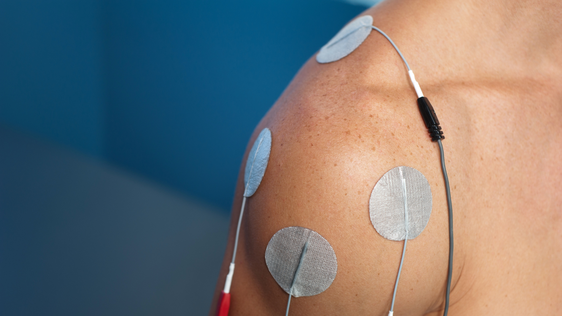
Neuromuscular electrical stimulation (NMES) is often associated with postoperative rehabilitation. However, growing evidence suggests that introducing NMES before surgery can play a meaningful role in improving patient outcomes (Punnoose et al., 2023; Walls et al., 2010; Patanè et al., 2025). This approach is based on a simple principle: the patient’s muscular status at the time of surgery influences postoperative recovery (Gillis et al., 2022; Hiyama et al., 2026). In other words, better preoperative muscle function is generally associated with better functional outcomes after surgery (Punnoose et al., 2023; Devasenapathy et al., 2019). This concept, often summarized as Better In, Better Out, is now supported by robust evidence across several orthopedic settings (Gillis et al., 2022; Jørgensen et al., 2024). Preoperative Muscle Strength: A Modifiable Prognostic Factor Among the many factors that influence recovery after orthopedic surgery, preoperative muscle strength stands out for two reasons. It is strongly associated with postoperative outcomes (Devasenapathy et al., 2019; Takamura et al., 2024; Hiyama et al., 2026), and it can be improved before surgery (Punnoose et al., 2023; Walls et al., 2010). Total Knee Arthroplasty In patients undergoing total knee arthroplasty, preoperative quadriceps strength is one of the strongest predictors of postoperative function (Devasenapathy et al., 2019; Hiyama et al., 2026). Patients with greater strength before surgery are more likely to achieve satisfactory mobility and functional recovery. Several studies have identified strength thresholds that predict walking ability six months after surgery (Takamura et al., 2024). More recently, Hiyama and colleagues (2026) reported that patients who regain age-adjusted normative quadriceps strength experience greater satisfaction and better functional outcomes one year after surgery. Anterior Cruciate Ligament Reconstruction A similar pattern is seen after anterior cruciate ligament reconstruction. Significant preoperative quadriceps weakness is associated with poorer knee function one year after surgery (Kim et al., 2022). Conversely, patients with greater preoperative strength tend to achieve better functional outcomes. These findings are consistent with the conclusions of a recent systematic review (Qiu et al., 2020). Shoulder Surgery Preoperative muscle strength also plays an important role in shoulder surgery. In patients undergoing anatomic or reverse shoulder arthroplasty, abduction and external rotation strength are among the strongest predictors of postoperative function (Hao et al., 2022a; Hao et al., 2022b). Grip strength, often considered a global indicator of muscle health and sarcopenia, has also been associated with outcomes following shoulder arthroplasty (Lee et al., 2023). More recent evidence suggests that it may also predict functional recovery after rotator cuff repair. NMES: An Opportunity Before Surgery If muscle strength affects postoperative outcomes, an obvious question arises: can this factor be optimized before surgery? NMES represents an attractive option, particularly for patients with pain, marked weakness, or functional limitations that restrict conventional exercise. In a pilot study involving patients awaiting total knee arthroplasty, a home-based NMES program increased quadriceps strength by approximately 28% before surgery (Walls et al., 2010). These gains translated into faster recovery of strength and function in the early postoperative period, while also limiting muscle atrophy. These findings align with the broader evidence supporting prehabilitation. A meta-analysis published in JAMA Network Open, which included 48 randomized controlled trials and more than 3,500 participants, found that preoperative interventions significantly improved muscle strength and physical function before surgery. Benefits persisted during the first weeks of postoperative recovery (Punnoose et al., 2023). The Role of NMES After Surgery NMES remains a valuable tool after surgery. During the early postoperative period, muscle recovery is limited by pain, joint effusion, immobilization, arthrogenic muscle inhibition, and reduced physical activity. In this context, NMES can effectively stimulate muscle activation despite these limitations. Following Knee Surgery After arthroplasty or ligament reconstruction, rapid quadriceps weakness represents a major clinical challenge. Early use of NMES may help attenuate strength loss and accelerate functional recovery, particularly when voluntary activation remains impaired. Following Shoulder Surgery Although evidence of preoperative NMES in shoulder surgery remains limited, postoperative findings are encouraging. Recent studies have shown that NMES may reduce deltoid atrophy after rotator cuff repair and accelerate recovery of abduction strength (Yoon et al., 2026). Other studies have reported earlier improvements in external rotation strength when NMES is applied to the infraspinatus during the initial phases of rehabilitation (Reinold et al., 2008). Sarcopenia: An Often Overlooked Factor Beyond muscle strength, preoperative muscle mass also appears to influence outcomes. A meta-analysis including more than 14,000 patients found that low preoperative muscle mass was associated with a significantly higher risk of major postoperative complications and short-term mortality (Weerink et al., 2020). In total knee arthroplasty, low muscle mass has also been identified as an independent risk factor for postoperative blood transfusion (Hwang et al., 2022). These findings highlight that preoperative preparation is not solely aimed at improving function. It may also help reduce surgical risk. Key Takeaways Current evidence points to a consistent conclusion: preoperative muscle status has a direct impact on recovery following orthopedic surgery. For rehabilitation professionals, this represents an opportunity to intervene before the operation takes place. NMES appears particularly valuable for optimizing a modifiable prognostic factor, especially when pain or functional limitations prevent patients from performing high-intensity strengthening exercises. From this perspective, the period leading up to surgery should no longer be viewed as passively waiting. Instead, it should be considered a therapeutic window during which clinicians can improve a patient’s muscle reserve, support postoperative recovery, and ultimately enhance functional outcomes. In orthopedic rehabilitation, recovery often begins long before the patient enters the operating room. References Bodkin, S. G., Norte, G. E., & Hart, J. M. (2019). Corticospinal excitability can discriminate quadriceps strength indicative of knee function after ACL-reconstruction. Scandinavian Journal of Medicine & Science in Sports, 29(5), 716–724. Devasenapathy, N., Maddison, R., Malhotra, R., Zodepy, S., & Sharma, S. (2019). Preoperative quadriceps muscle strength and functional ability predict performance-based outcomes 6 months after total knee arthroplasty: A systematic review. Physical Therapy, 99(1), 46–61. Gillis, C., Ljungqvist, O., & Carli, F. (2022). Prehabilitation, enhanced recovery after surgery, or both? A narrative review. British Journal of Anaesthesia, 128(3), 434–448. Hao, K. A., Wright, T. W., Schoch, B. S., Struk, A. M., King, J. J., & Farmer, K. W. (2022a). Association between preoperative shoulder strength and clinical outcomes after primary reverse total shoulder arthroplasty. Journal of the American Academy of Orthopaedic Surgeons, 30(15), e1035–e1044. Hao, K. A., Wright, T. W., Dean, E. W., Struk, A. M., & King, J. J. (2022b). Preoperative shoulder strength is associated with postoperative primary anatomic total shoulder arthroplasty outcomes and improvement. Journal of Shoulder and Elbow Surgery, 31(8), 1648–1656. Hauger, A. V., Reiman, M. P., Bjordal, J. M., Sheets, C., Ledbetter, L., & Goode, A. P. (2018). Neuromuscular electrical stimulation is effective in strengthening the quadriceps muscle after anterior cruciate ligament surgery. Knee Surgery, Sports Traumatology, Arthroscopy, 26(2), 399–410. Hiyama, Y., Sawa, R., Yokoyama, M., & Ishii, Y. (2026). Achieving normative quadriceps strength after total knee arthroplasty: Associations with satisfaction, function, and a predictive nomogram. The Bone & Joint Journal, 108-B(1), 45–52. Hwang, D., Han, H. S., Lee, M. C., & Ro, D. H. (2022). Low muscle mass is an independent risk factor for postoperative blood transfusion in total knee arthroplasty: A retrospective, propensity score-matched cohort study. BMC Geriatrics, 22(1), Article 137. Jenssen, K. K., Lundgreen, K., Madsen, J. E., Kvakestad, R., & Dimmen, S. (2018). Prognostic factors for functional outcome after rotator cuff repair: A prospective cohort study with 2-year follow-up. The American Journal of Sports Medicine, 46(14), 3504–3510. Jørgensen, S. L., Aagaard, P., Bohn, M. B., Mikkelsen, L. R., & Mechlenburg, I. (2024). The effect of blood flow restriction exercise prior to total knee arthroplasty on postoperative physical function, lower limb strength and patient-reported outcomes: A randomized controlled trial. Scandinavian Journal of Medicine & Science in Sports, 34(1), e14516. Kim, D. K., Park, G., Wang, J. H., Kuo, L. T., & Park, W. H. (2022). Preoperative quadriceps muscle strength deficit severity predicts knee function one year after anterior cruciate ligament reconstruction. Scientific Reports, 12(1), Article 11845. Lee, B. G., Lee, D., & Koh, J. H. (2023). Relationship between the preoperative grip strength and postoperative shoulder strength of patients treated via reverse shoulder arthroplasty. Journal of Shoulder and Elbow Surgery, 32(5), 1012–1019. Patanè, P., Carnevale Pellino, V., Febbi, M., Gatti, R., & Sirtori, V. (2025). Effects of a tele-prehabilitation program with indirect electrostimulation compared to home-based exercise in patients eligible for lower limb arthroplasty: A randomized controlled trial. Journal of Clinical Medicine, 14(3), Article 892. Punnoose, A., Claydon-Mueller, L. S., Weiss, O., Zhang, J., Maffulli, N., & Rushton, A. (2023). Takamura, D., Iwata, K., Yajima, Y., et al. (2024). Cut-off values of preoperative knee extensor strength and hip abductor strength for predicting good walking ability after total knee arthroplasty. Archives of Orthopaedic and Trauma Surgery, 144(1), 305–312. Walls, R. J., McHugh, G., O'Gorman, D. J., Moyna, N. M., & O'Byrne, J. M. (2010). Effects of preoperative neuromuscular electrical stimulation on quadriceps strength and functional recovery in total knee arthroplasty: A pilot study. BMC Musculoskeletal Disorders, 11, 119.
On associe souvent la stimulation électrique neuromusculaire (NMES) à la phase postopératoire. Cependant, des preuves de plus en plus convaincantes indiquent que son utilisation précoce, avant l’intervention chirurgicale, peut contribuer significativement à la réadaptation des patients (Punnoose et al., 2023 ; Walls et al., 2010 ; Patanè et al., 2025). Cette évolution repose sur une idée simple : l'état musculaire dans lequel un patient entre en chirurgie influence directement sa capacité à récupérer par la suite (Gillis et al., 2022 ; Hiyama et al., 2026). En d'autres termes, une meilleure condition musculaire préopératoire est généralement associée à de meilleurs résultats fonctionnels postopératoires (Punnoose et al., 2023 ; Devasenapathy et al., 2019). Ce principe, parfois résumé par l'expression Better In, Better Out, est aujourd'hui soutenu par des données robustes dans plusieurs contextes orthopédiques (Gillis et al., 2022 ; Jørgensen et al., 2024). La force musculaire préopératoire : un facteur pronostique modifiable Parmi les nombreux facteurs susceptibles d'influencer la récupération après une chirurgie orthopédique, la force musculaire préopératoire se distingue par deux caractéristiques importantes : elle est fortement associée aux résultats postopératoires (Devasenapathy et al., 2019 ; Takamura et al., 2024 ; Hiyama et al., 2026) et elle peut être améliorée avant l'intervention. (Punnoose et al., 2023 ; Walls et al., 2010) Arthroplastie totale du genou Chez les personnes qui subissent une arthroplastie totale du genou, la force du quadriceps avant l'opération est l'un des meilleurs prédicteurs de la récupération fonctionnelle ultérieure (Devasenapathy et al., 2019; Hiyama et al., 2026). Les patients présentant une meilleure force musculaire avant la chirurgie ont davantage de chances d'atteindre des niveaux satisfaisants de fonction et de mobilité après l'intervention. Certaines études ont même identifié des seuils de force permettant de prédire la capacité de marche à six mois postopératoires (Takamura et al., 2024). Plus récemment, Hiyama et ses collègues (2026) ont montré que les patients qui retrouvent une force du quadriceps comparable aux valeurs normatives rapportent une meilleure satisfaction et de meilleurs résultats fonctionnels un an après leur chirurgie. Reconstruction du ligament croisé antérieur La même tendance est observée après une reconstruction du ligament croisé antérieur. Un déficit préopératoire important de force du quadriceps est associé à une moins bonne fonction du genou à un an (Kim et al., 2022). À l'inverse, les patients qui présentent une meilleure force musculaire avant l'intervention obtiennent généralement de meilleurs résultats fonctionnels postopératoires. Cette association est également appuyée par les conclusions d'une revue systématique récente (Qiu et al., 2020). Chirurgies de l'épaule À l'épaule, la force musculaire préopératoire joue également un rôle déterminant. Chez les patients qui subissent une arthroplastie anatomique ou inversée, la force en abduction et en rotation externe figure parmi les prédicteurs les plus puissants des résultats fonctionnels postopératoires (Hao et al., 2022a; Hao et al., 2022b). La force de préhension, souvent considérée comme un indicateur global de l'état musculaire et de la sarcopénie, s'avère également associée aux résultats postopératoires après arthroplastie de l'épaule (Lee et al., 2023). Plus récemment, des données suggèrent qu'elle pourrait aussi prédire la récupération fonctionnelle après réparation de la coiffe des rotateurs. La SNME : une occasion d'agir avant la chirurgie Si la force musculaire influence le pronostic postopératoire, la question devient alors évidente : peut-on améliorer ce facteur avant l'intervention? La NMES constitue une avenue particulièrement intéressante pour y parvenir, notamment chez les patients qui présentent de la douleur, une faiblesse importante ou des limitations fonctionnelles qui restreignent leur capacité à s'entraîner de façon conventionnelle. Dans une étude pilote réalisée auprès de patients en attente d'une arthroplastie totale du genou, un programme de NMES à domicile a permis d'augmenter la force du quadriceps d'environ 28 % avant la chirurgie (Walls et al., 2010). Ces gains se sont traduits par une récupération plus rapide de la force et de la fonction durant les semaines suivant l'intervention, tout en limitant l'atrophie musculaire postopératoire. Ces résultats s'inscrivent dans un ensemble de données plus large soutenant les bénéfices de la préhabilitation. Une méta-analyse publiée dans JAMA Network Open, regroupant 48 essais cliniques et plus de 3 500 participants, a démontré que les interventions réalisées avant la chirurgie améliorent significativement la force musculaire et la fonction préopératoires, avec des bénéfices qui se maintiennent durant les premières semaines de récupération postopératoire (Punnoose et al., 2023). Le rôle de la SNME après la chirurgie La NMES demeure toutefois un outil pertinent après l'intervention. Au cours des premières semaines postopératoires, plusieurs facteurs limitent la récupération musculaire : douleur, épanchement articulaire, immobilisation, inhibition musculaire réflexe et réduction de l'activité physique. Dans ce contexte, la NMES permet de solliciter efficacement le muscle malgré les limitations fonctionnelles présentes. Après une chirurgie du genou Après une arthroplastie ou une reconstruction ligamentaire, la perte rapide de force du quadriceps représente un défi clinique majeur. L'utilisation précoce de la NMES peut contribuer à atténuer cette perte et favoriser une récupération fonctionnelle plus rapide, particulièrement lorsque l'activation volontaire demeure insuffisante. Après une chirurgie de l'épaule Bien que les données sur la NMES préopératoire à l'épaule soient encore limitées, les résultats postopératoires sont prometteurs. Des études récentes démontrent que la NMES peut réduire l'atrophie du deltoïde après une réparation de la coiffe des rotateurs et accélérer la récupération de la force en abduction (Yoon et al., 2026). D'autres travaux ont également observé une amélioration précoce de la force en rotation externe lorsque la NMES est appliquée à l'infraépineux durant les premières phases de réadaptation (Reinold et al., 2008). La sarcopénie : un facteur souvent sous-estimé Au-delà de la force musculaire, la quantité de masse musculaire disponible avant la chirurgie semble également influencer les résultats. Une méta-analyse regroupant plus de 14 000 patients a montré qu'une faible masse musculaire préopératoire est associée à une augmentation significative du risque de complications postopératoires majeures et de mortalité à court terme (Weerink et al., 2020). En arthroplastie du genou, une faible masse musculaire a également été identifiée comme un facteur de risque indépendant de transfusion postopératoire (Hwang et al., 2022). Ces observations rappellent que la préparation préopératoire ne vise pas uniquement à améliorer la fonction. Elle peut également contribuer à réduire certains risques associés à l'intervention chirurgicale. Ce qu'il faut retenir Les données actuelles convergent vers un même constat : la condition musculaire préopératoire influence directement la récupération après une chirurgie orthopédique. Pour les professionnels de la réadaptation, cela représente une occasion importante d'agir avant même que l'intervention n'ait lieu. La NMES apparaît comme une stratégie particulièrement intéressante pour optimiser un facteur pronostique modifiable, surtout lorsque la douleur ou les limitations fonctionnelles compromettent la capacité du patient à réaliser un entraînement musculaire suffisamment intense. Dans cette perspective, la période précédant la chirurgie ne devrait plus être considérée comme une simple attente. Elle constitue plutôt une fenêtre thérapeutique où il est possible d'améliorer la réserve musculaire du patient, de favoriser sa récupération future et, ultimement, d'optimiser ses résultats fonctionnels. Parce qu'en réadaptation orthopédique, la récupération commence souvent bien avant l'entrée en salle d'opération. Bodkin, S. G., Norte, G. E., & Hart, J. M. (2019). Corticospinal excitability can discriminate quadriceps strength indicative of knee function after ACL-reconstruction. Scandinavian Journal of Medicine & Science in Sports, 29(5), 716–724. Devasenapathy, N., Maddison, R., Malhotra, R., Zodepy, S., & Sharma, S. (2019). Preoperative quadriceps muscle strength and functional ability predict performance-based outcomes 6 months after total knee arthroplasty: A systematic review. Physical Therapy, 99(1), 46–61. Gillis, C., Ljungqvist, O., & Carli, F. (2022). Prehabilitation, enhanced recovery after surgery, or both? A narrative review. British Journal of Anaesthesia, 128(3), 434–448. Hao, K. A., Wright, T. W., Schoch, B. S., Struk, A. M., King, J. J., & Farmer, K. W. (2022a). Association between preoperative shoulder strength and clinical outcomes after primary reverse total shoulder arthroplasty. Journal of the American Academy of Orthopaedic Surgeons, 30(15), e1035–e1044. Hao, K. A., Wright, T. W., Dean, E. W., Struk, A. M., & King, J. J. (2022b). Preoperative shoulder strength is associated with postoperative primary anatomic total shoulder arthroplasty outcomes and improvement. Journal of Shoulder and Elbow Surgery, 31(8), 1648–1656. Hauger, A. V., Reiman, M. P., Bjordal, J. M., Sheets, C., Ledbetter, L., & Goode, A. P. (2018). Neuromuscular electrical stimulation is effective in strengthening the quadriceps muscle after anterior cruciate ligament surgery. Knee Surgery, Sports Traumatology, Arthroscopy, 26(2), 399–410. Hiyama, Y., Sawa, R., Yokoyama, M., & Ishii, Y. (2026). Achieving normative quadriceps strength after total knee arthroplasty: Associations with satisfaction, function, and a predictive nomogram. The Bone & Joint Journal, 108-B(1), 45–52. Hwang, D., Han, H. S., Lee, M. C., & Ro, D. H. (2022). Low muscle mass is an independent risk factor for postoperative blood transfusion in total knee arthroplasty: A retrospective, propensity score-matched cohort study. BMC Geriatrics, 22(1), Article 137. Jenssen, K. K., Lundgreen, K., Madsen, J. E., Kvakestad, R., & Dimmen, S. (2018). Prognostic factors for functional outcome after rotator cuff repair: A prospective cohort study with 2-year follow-up. The American Journal of Sports Medicine, 46(14), 3504–3510. Jørgensen, S. L., Aagaard, P., Bohn, M. B., Mikkelsen, L. R., & Mechlenburg, I. (2024). The effect of blood flow restriction exercise prior to total knee arthroplasty on postoperative physical function, lower limb strength and patient-reported outcomes: A randomized controlled trial. Scandinavian Journal of Medicine & Science in Sports, 34(1), e14516. Kim, D. K., Park, G., Wang, J. H., Kuo, L. T., & Park, W. H. (2022). Preoperative quadriceps muscle strength deficit severity predicts knee function one year after anterior cruciate ligament reconstruction. Scientific Reports, 12(1), Article 11845. Lee, B. G., Lee, D., & Koh, J. H. (2023). Relationship between the preoperative grip strength and postoperative shoulder strength of patients treated via reverse shoulder arthroplasty. Journal of Shoulder and Elbow Surgery, 32(5), 1012–1019. Patanè, P., Carnevale Pellino, V., Febbi, M., Gatti, R., & Sirtori, V. (2025). Effects of a tele-prehabilitation program with indirect electrostimulation compared to home-based exercise in patients eligible for lower limb arthroplasty: A randomized controlled trial. Journal of Clinical Medicine, 14(3), Article 892. Punnoose, A., Claydon-Mueller, L. S., Weiss, O., Zhang, J., Maffulli, N., & Rushton, A. (2023). Takamura, D., Iwata, K., Yajima, Y., et al. (2024). Cut-off values of preoperative knee extensor strength and hip abductor strength for predicting good walking ability after total knee arthroplasty. Archives of Orthopaedic and Trauma Surgery, 144(1), 305–312. Walls, R. J., McHugh, G., O'Gorman, D. J., Moyna, N. M., & O'Byrne, J. M. (2010). Effects of preoperative neuromuscular electrical stimulation on quadriceps strength and functional recovery in total knee arthroplasty: A pilot study. BMC Musculoskeletal Disorders, 11, 119.